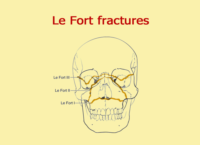
 softness and separation of the zygomatic-frontal suture. This type of fracture predisposes the patient more to cerebrospinal fluid rhinorrhoea than the other two. Within the nose, a branch of the fracture extends through the base of the perpendicular lamina of the ethmoid, through the vomer and into the pterygoid processes at the base of the sphenoid. The fracture then continues along the floor of the orbit, along the inferior orbital fissure and continues superiorly and laterally through the lateral wall of the orbit, through the zygomatic-frontal suture and the zygomatic arch. The thickness of the sphenoid posteriorly usually prevents continuation of the fracture into the optic canal. This fracture begins at the fronto-maxillary suture and the naso-frontal suture and extends posteriorly along the medial wall of the orbit through the nasolacrimal groove and the ethmoid. It can occur as a result of impact on the root of the nose or the upper part of the jawbone. LeFort III fracture, also called high, transverse or craniofacial disjunction, usually involves the zygomatic arch. LeFort III fracture (high, transverse fracture or craniofacial disjunction) anaesthesia or paresthesia of the cheek (from damage to the infraorbital nerve). This fracture is pyramidal in shape, extending from the root of the nose, at or just below the naso-frontal suture, through the frontal processes of the maxillary bone, then laterally and downward through the lacrimal bones and the lower floor of the orbit, reappearing through or near the infraorbital foramen and inferiorly through the anterior wall of the maxillary sinus it then proceeds below the zygomatic bone, through the pterygomaxillary fissure to terminate on the pterygoid processes of the sphenoid. LeFort II fracture, also called medium or pyramidal fracture, may result from trauma to the middle or lower jaw, and usually involves the lower edge of the orbit. LeFort II fracture (medium or pyramidal fracture) soft tissue oedema in the middle third of the face. Some symptoms may be present in both LeFort I and LeFort II, such as: Percussion of the upper jaw teeth reveals a sound known as a foul pot. LeFort I fractures may be almost immobile, and the characteristic screeching can only be perceived by applying pressure to the teeth of the upper arch. Guérin’s sign is present, characterised by ecchymosis in the region of the greater palatine vessels. ecchymosis in the upper fornix under the zygomatic arches,. The fracture extends from the nasal septum to the lateral edges of the piriform opening, travels horizontally above the tooth apices, crosses under the zygomatic-jaw suture and crosses the sphenoid-jaw suture to interrupt the pterygoid processes of the sphenoid. It is also known as Guérin’s fracture, or floating palate, and usually involves the lower portion of the pyriform opening. LeFort I fracture, also known as a low or horizontal fracture, can result from a downward force on the alveolar border of the maxilla. LeFort I fracture (low or horizontal fracture) The diagnosis of LeFort fractures is made through an objective examination (in which the palate is often unnaturally mobile) supported by a CT scan of the head and neck, which in most cases is able to clearly show the type of fracture.įor fractures to be classified as LeFort, they must involve the pterygoid processes of the sphenoid, which are visible posterior to the maxillary sinuses in an axial CT scan, and inferior to the orbital rim in a coronal projection.
fractures occurring in tissues affected by internal structural failure due to an underlying pathology that may be systemic or local. In this case we speak of pathological fractures, i.e. general factors: osteomalacia and osteopetrosis, hyperparathyroidism, senile osteoporosis, occupational phosphorus or fluoride toxicosis.local factors: non-specific and specific infectious processes, malignant and benign tumours, cysts, dental retention.

LeFort fractures can also be favoured by a variety of factors, such as LeFort fractures are most often caused by direct trauma to the face and head in general, for example in road accidents, and are often associated with a variety of other injuries to the rest of the body. Causes and risk factors of LeFort fractures posterior pillar (pterygomatic): from the tuberosity of the maxilla it leads to the pterygoid processes of the sphenoid bone.įracture lines in facial trauma tend to occur at the periphery of the areas traversed by these trajectories, resulting in the different types of LeFort fracture.lateral pillar (zygomatic): from the molar region it follows the lateral wall of the orbit.anterior (naso-frontal) pillar: starts at the piriform opening and follows the medial orbital frame, surrounding the canine region inferiorly.


 0 kommentar(er)
